
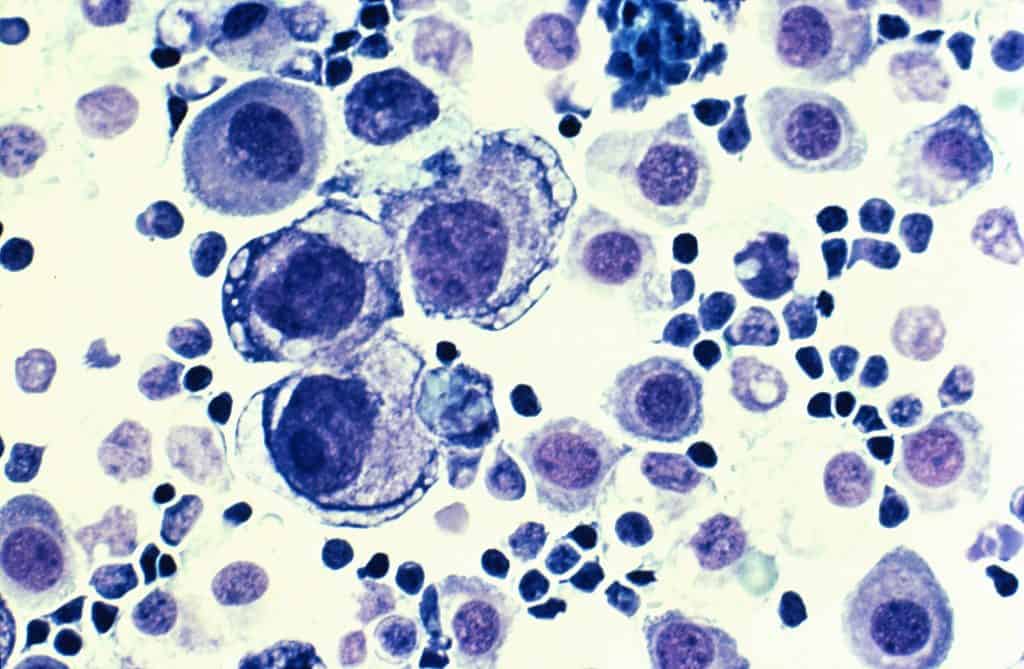
Introduction to PML and PML Nuclear Bodies
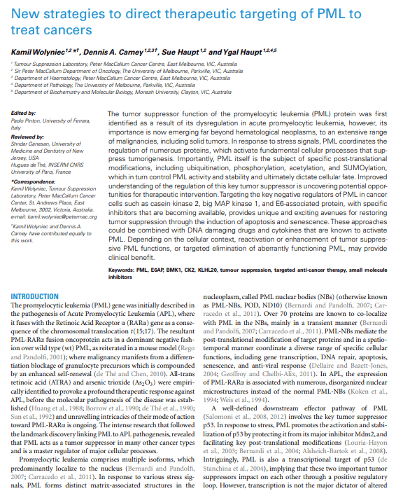
The promyelocytic leukemia (PML) protein is a multifunctional tumor suppressor that plays a central role in regulating apoptosis, genomic stability, transcriptional control, DNA repair, and cellular senescence. The PML gene spans nearly 53 kb within the human genome and undergoes extensive alternative splicing, generating multiple protein isoforms with distinct biological functions. Experimental studies have identified six nuclear isoforms and one cytoplasmic variant. Among these, PML I represents the longest isoform, whereas PML VII is the shortest.
Despite differences in their C-terminal regions, all PML isoforms share a highly conserved N-terminal domain composed of several structural motifs, including:
- A RING finger domain
- Two cysteine/histidine-rich B-box domains
- A coiled-coil region
Together, these motifs form the RBCC or TRIM domain, which is essential for protein-protein interactions, oligomerization, and nuclear body assembly. The variable C-terminal sequences confer isoform-specific activities, enabling different PML variants to participate in specialized signaling pathways and transcriptional programs.
One important example involves PML II, which is required for interferon-mediated activation of immune-responsive genes through interactions with transcription factors such as NF-κB, STAT1, and CBP. Certain isoforms, including PML II and PML V, can also generate nuclear bodies independently of the classical RBCC domain, highlighting the structural complexity of PML regulation.
PML Nuclear Bodies: Dynamic Regulatory Centers in the Nucleus
PML proteins are predominantly concentrated within specialized punctate structures dispersed throughout the nucleus, commonly known as PML nuclear bodies (PML-NBs). These structures are also referred to as:
- Nuclear domain 10 (ND10)
- PODs (PML oncogenic domains)
- Kremer bodies
PML-NBs are highly dynamic macromolecular assemblies that vary in size, number, and composition depending on cellular stress, nutrient availability, cell cycle phase, and cell type. Typically, a nucleus contains between 5 and 30 PML bodies.
The RBCC domain is indispensable for the formation and structural organization of these nuclear compartments. Loss of PML expression leads to complete disruption of PML-NBs, demonstrating that PML functions as their architectural scaffold.
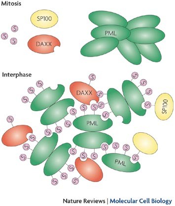
Mechanisms of PML-NB Formation
Two major models have been proposed to explain PML nuclear body assembly:
1. SUMO-Dependent Assembly Model
This model suggests that PML sumoylation and SUMO-SIM interactions initiate nuclear body nucleation. PML proteins contain SUMO-interacting motifs (SIMs) that promote non-covalent interactions between SUMOylated proteins and PML molecules.
2. Oxidative Stress-Driven Model
An alternative model proposes a two-step process in which reactive oxygen species (ROS) first induce oxidation-mediated covalent interactions between PML monomers. Subsequent recruitment of SUMOylated proteins and SIM-containing partners then enhances maturation of the nuclear body.
This second model helps explain why certain PML mutants lacking SUMOylation sites can still form nuclear aggregates.
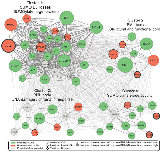
Protein Interaction Network of PML
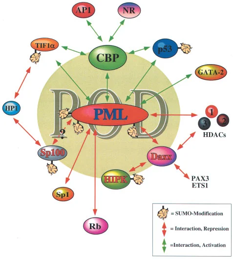
PML functions as a major signaling hub within the nucleus. More than 160 proteins are known to interact directly or indirectly with PML, including:
- p53
- Mdm2
- Akt
- DAXX
- STAT proteins
- DNA repair enzymes
- Chromatin remodelers
These interactions allow PML to coordinate multiple cellular pathways simultaneously, particularly those associated with tumor suppression and stress responses.
Tumor Suppressive Functions of PML
PML was identified as a tumor suppressor shortly after discovery of the PML gene. Reduced PML expression has since been observed in numerous malignancies, including:
- Breast cancer
- Lung cancer
- Colorectal carcinoma
- Prostate cancer
- Bladder cancer
Experimental overexpression of PML suppresses cellular proliferation and promotes:
- Cell cycle arrest
- Senescence
- Apoptosis
Conversely, loss of PML enhances resistance to apoptosis and increases tumor susceptibility in animal models.
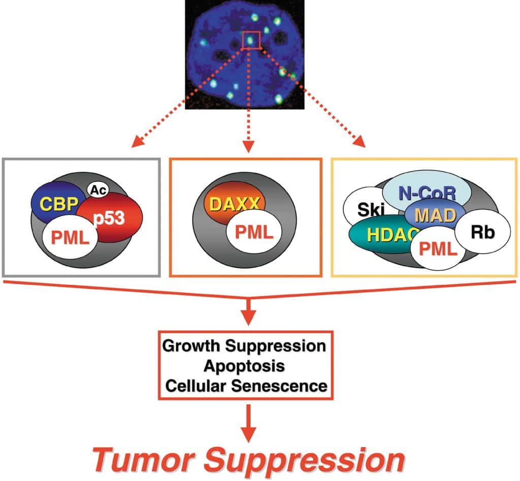
Functional Mechanisms of Tumor Suppression
PML exerts antitumor activity through several interconnected mechanisms:
- Sequestration of regulatory proteins within PML-NBs
- Promotion of protein-protein interactions
- Regulation of post-translational modifications
- Modulation of transcription factor activity
- Facilitation of DNA damage repair
- Maintenance of telomere integrity through ALT pathways
These processes collectively regulate critical pathways controlling cell survival, genomic integrity, and proliferation.
PML and Apoptosis Regulation
Apoptosis is a major anticancer mechanism controlled by PML. The protein promotes both caspase-dependent and caspase-independent cell death pathways.
Caspase-3 Activation
PML stimulates activation of caspase-3 following exposure to:
- Gamma irradiation
- TNFα
- Fas signaling
- Interferons
- Ceramide
Cells lacking PML display resistance to apoptotic stimuli, demonstrating the importance of PML-mediated caspase activation in tumor suppression.
Caspase-Independent Apoptosis
PML can also induce apoptosis independently of caspase-3 by recruiting pro-apoptotic proteins such as:
- BAX
- p27KIP1
to PML nuclear bodies.
PML as a Master Regulator of p53 Activity
The tumor suppressor p53 is one of the most important targets regulated by PML.
Stabilization of p53
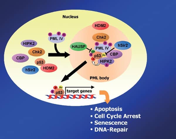
Under stress conditions, PML enhances p53 stability by sequestering the E3 ubiquitin ligase Mdm2 inside PML-NBs, thereby preventing p53 degradation.
PML also promotes recruitment of:
- Chk2 kinase
- CBP/p300 acetyltransferases
- HIPK2 kinase
These enzymes modify p53 through phosphorylation and acetylation, enhancing its transcriptional activity and promoting apoptosis or senescence.
Negative Regulation via SIRT1
PML-NBs can also recruit the deacetylase SIRT1, which suppresses p53 transcriptional activity through deacetylation. Thus, the biological outcome depends on the specific composition of proteins assembled within the nuclear body.
Inhibition of the Akt Survival Pathway
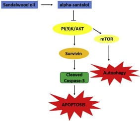
Akt signaling promotes cell growth, survival, and metabolic activity in many cancers. PML negatively regulates this oncogenic pathway through several mechanisms.
PP2A-Mediated Akt Inactivation
PML recruits protein phosphatase 2A (PP2A) into nuclear bodies, where PP2A dephosphorylates Akt and inhibits its kinase activity.
Regulation of PTEN and eIF4E
PML additionally suppresses Akt signaling by:
- Inhibiting eIF4E-mediated mRNA export
- Stabilizing nuclear PTEN
- Blocking HAUSP-mediated PTEN deubiquitination
Through these mechanisms, PML suppresses cell proliferation and promotes apoptosis.
PML and Retinoblastoma Protein (RB) Regulation
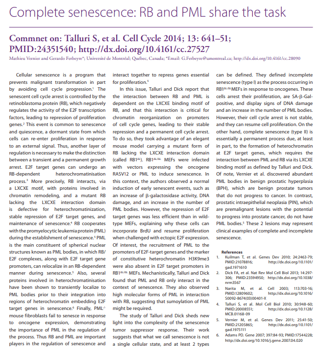
The RB protein suppresses cell cycle progression by inhibiting E2F transcription factors. PML-NBs recruit phosphatase PP1α, which dephosphorylates RB and restores its inhibitory interaction with E2F.
This mechanism contributes to:
- Cell cycle arrest
- Cellular senescence
- Suppression of uncontrolled proliferation
Transcriptional Regulation by PML
PML functions as both a transcriptional repressor and activator depending on cellular context.
Transcriptional Repression
PML suppresses gene expression by sequestering transcription factors such as:
- NF-κB
- STAT3
- Sp1
- Nur77
within nuclear bodies, preventing their access to target promoters.
PML also forms complexes with transcriptional corepressors and histone deacetylases to inhibit oncogenic transcriptional programs.
Transcriptional Activation
In contrast, PML can enhance transcription by:
- Stabilizing transcription factors
- Sequestering transcriptional repressors
- Facilitating assembly of transactivation complexes
PML positively regulates pathways involving:
- Interferon signaling
- β-catenin-responsive genes
- GATA transcription factors
- AP-1 signaling
- p21WAF1/CIP1 activation
These dual activities illustrate the versatility of PML in gene regulation.
PML in DNA Damage Repair and Genome Stability
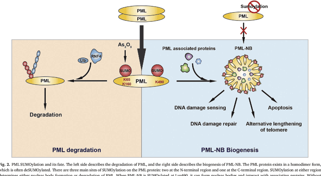
Emerging evidence demonstrates that PML is critically involved in DNA repair pathways and telomere maintenance.
Alternative Lengthening of Telomeres (ALT)
In telomerase-negative cancer cells, PML-NBs associate with:
- Telomeric DNA
- TRF1/TRF2 proteins
- RAD51
- NBS1
- Mre11
forming specialized ALT-associated PML bodies that maintain telomere integrity.
Double-Strand Break Repair
PML also participates in repair of DNA double-strand breaks by coordinating recruitment of repair proteins including:
- ATM
- ATR
- Chk2
- RAD50-Mre11-NBS1 complex
- BLM helicase
These functions are essential for preserving genomic stability and preventing tumorigenesis.
Cytoplasmic PML: Tumor Suppressor or Oncoprotein?
Although PML is classically nuclear, several cytoplasmic isoforms have been identified with context-dependent functions.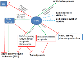
Oncogenic Functions
Certain truncated cytoplasmic PML variants can:
- Sequester nuclear PML
- Inhibit p53 activity
- Promote resistance to apoptosis
- Enhance tumor cell proliferation
These forms are frequently associated with aggressive acute promyelocytic leukemia (APL).
Tumor Suppressive Functions
Conversely, cytoplasmic PML can suppress tumor growth by:
- Inhibiting PKM2 metabolic activity
- Regulating TGFβ signaling
- Promoting calcium-mediated apoptosis through ER-mitochondrial signaling
Thus, cytoplasmic PML exhibits dual biological roles depending on cellular context and molecular interactions.
Regulation of PML Expression
PML expression is tightly regulated at multiple levels.
Transcriptional Regulation
Inflammatory cytokines strongly induce PML transcription, including:
- Interferons
- TNFα
- IL-6
These effects are mediated primarily through JAK/STAT and NF-κB signaling pathways.
Translational Regulation
PML mRNA translation can be enhanced through:
- mTOR signaling
- eIF4E activity
- Internal ribosome entry site (IRES)-dependent mechanisms
particularly during stress responses.
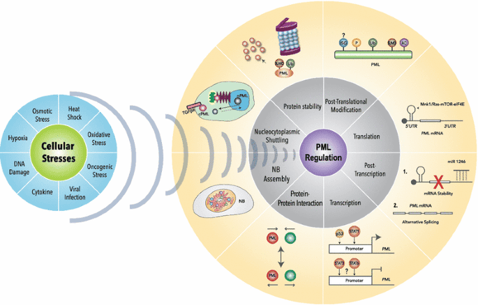
Post-Translational Regulation of PML
PML protein abundance and activity are controlled by extensive post-translational modifications.
Major Regulatory Modifications
These include:
- SUMOylation
- Ubiquitination
- Phosphorylation
- Acetylation
- Isomerization
Cross-talk among these modifications determines PML stability, localization, and activity.
Ubiquitin-Mediated Degradation
Several E3 ligases target PML for degradation, including:
- RNF4
- KLHL20
- UHRF1
- E6AP
- SIAH proteins
By contrast, USP11 stabilizes PML through deubiquitination.
SUMOylation and Arsenic Trioxide
SUMOylation is critical for PML-NB formation and protein recruitment. Therapeutically, arsenic trioxide promotes SUMO-dependent degradation of the oncogenic PML-RARα fusion protein in acute promyelocytic leukemia.
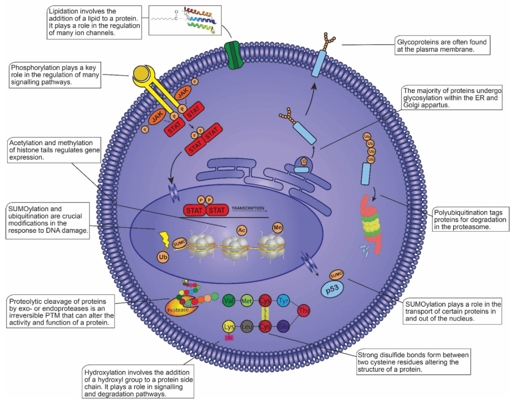
Nucleocytoplasmic Shuttling of PML
PML localization between the nucleus and cytoplasm is dynamically regulated.
Oxidative stress, acetylation status, and antioxidant signaling can alter PML trafficking. For instance:
- Hydrogen peroxide promotes nuclear accumulation
- Sulforaphane induces cytoplasmic redistribution
These localization changes significantly influence apoptosis, angiogenesis, and stress responses.
Therapeutic Potential of Targeting PML
Restoring or enhancing PML function represents an attractive anticancer strategy.
Several compounds increase PML abundance or activity, including:
- Arsenic trioxide
- Interferons
- Sulforaphane
- HDAC inhibitors
- Proteasome inhibitors
- CK2 inhibitors
- BMK1 inhibitors
Combination therapies that simultaneously stabilize PML and activate tumor suppressive pathways may provide improved anticancer efficacy.
PML in Angiogenesis and Tumor Microenvironment
PML suppresses tumor angiogenesis by inhibiting:
- mTOR signaling
- HIF-1α translation
- Integrin β1 expression
PML is also required for anti-angiogenic responses mediated by TNFα, interferons, and sulforaphane.
These findings highlight the broader role of PML beyond tumor cells themselves, extending into regulation of the tumor microenvironment.
Future Perspectives in PML Research
Emerging studies indicate that PML may play crucial roles in:
- Epigenetic regulation
- Chromatin remodeling
- Stem cell biology
- Metabolic control
- Brain development
PML interacts with multiple chromatin-modifying enzymes including:
- HDACs
- EZH2
- SETDB1
- UHRF1
- Histone acetyltransferases
However, the precise epigenetic mechanisms governed by PML remain poorly understood.
Another major challenge is explaining why PML acts as a tumor suppressor in some tissues but may exhibit oncogenic activity in others, such as chronic myeloid leukemia and triple-negative breast cancer.
Understanding these context-specific functions will be essential for developing precision therapies targeting the PML signaling network.
Conclusion
PML is a highly versatile tumor suppressor protein that coordinates multiple cellular defense mechanisms against malignant transformation. Through its organization of PML nuclear bodies, regulation of apoptosis, control of p53 and Akt signaling, participation in DNA repair, and modulation of transcriptional programs, PML serves as a central regulator of cellular homeostasis and genome integrity.
The complexity of PML regulation including its alternative splicing, post-translational modifications, and dynamic subcellular localization reflects its broad biological significance. Continued investigation into PML biology may uncover novel therapeutic strategies capable of restoring tumor suppressive functions and improving cancer treatment outcomes across multiple malignancies.