
Introduction to Experimental Retinoblastoma Models
Retinoblastoma is a rare but highly aggressive malignant retinal tumor that primarily affects children. Over the years, researchers have attempted to reproduce retinoblastoma experimentally in laboratory animals in order to better understand retinal oncogenesis, tumor differentiation, and mechanisms of viral or chemical carcinogenesis. Early experimental studies failed to generate true malignant retinal tumors despite inducing retinal hyperplasia or other ocular lesions.
For example, intraocular injections of heated lipids such as olive oil or lard into rat vitreous chambers produced retinal cell proliferation but did not lead to malignant transformation of sensory retinal tissue. Similarly, implantation of carcinogenic methylcholanthrene pellets in mouse eyes generated melanomas and squamous cell carcinomas rather than authentic retinal tumors. These observations highlighted the difficulty of experimentally inducing tumors that genuinely resemble human retinoblastoma.
The discovery that Human Adenovirus Type 12 (Ad12) could induce neurogenic tumors in the peripheral and central nervous systems of rodents opened new perspectives in retinal cancer research. Previous investigations demonstrated that Ad12 possesses strong oncogenic activity toward immature neuronal precursor cells, especially during early developmental stages. This suggested that undifferentiated retinal neuroblasts in newborn rats might represent susceptible targets for viral oncogenesis.
At the same time, chemical carcinogens such as N-methyl-N-nitrosourea (MNU) and ethylnitrosourea had already been shown to induce various neurogenic tumors when administered during fetal development. These findings emphasized the extreme sensitivity of immature neural tissues to carcinogenic transformation. Researchers therefore hypothesized that combining prenatal exposure to MNU with postnatal Ad12 inoculation might enhance retinal tumor development and reduce tumor latency.

Materials and Experimental Methods
Viral Preparation and Titration
The Huie strain of Ad12 was propagated in cultured HeLa cells under controlled laboratory conditions. Viral titration was performed using serial dilutions in primary human embryonic kidney cell cultures. Cytopathic effects were monitored for two weeks, allowing determination of viral infectivity levels. The final viral suspension reached a concentration of approximately 10⁸ tissue culture infective doses per milliliter, ensuring sufficient oncogenic potential for experimental inoculation.
Animal Experimental Design
Pregnant Sprague-Dawley CD rats were divided into multiple experimental groups to evaluate the isolated and combined effects of viral infection and chemical carcinogenesis.
Group Organization
- Group 1: Newborn rats received intraocular injections of Ad12 without MNU pretreatment.
- Group 2: Newborn rats were prenatally exposed to MNU and subsequently inoculated with Ad12 after birth.
- Group 3: Control animals received prenatal MNU exposure but no viral inoculation.
- Group 4: Control animals received neither MNU pretreatment nor active virus.
The virus or control fluid was injected directly into the left eye within 24 hours after birth. MNU was freshly prepared immediately before administration to preserve carcinogenic activity.
Animals that failed to develop tumors were sacrificed approximately 350 days after inoculation, and ocular tissues were processed for histopathological examination.
Histological and Ultrastructural Analysis
Histopathology
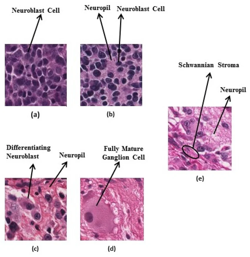
Tumor-bearing animals underwent perfusion fixation before tissue collection. Ocular tumors were embedded in paraffin, sectioned, and stained using multiple histological techniques, including:
- Hematoxylin-eosin staining
- Phosphotungstic acid hematoxylin
- Holmes silver impregnation
- Nissl staining
- Toluidine blue staining
These methods allowed detailed evaluation of tumor morphology, neuronal differentiation, and tissue architecture.
Electron Microscopy
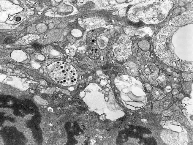
Ultrastructural examination was performed using transmission electron microscopy. Tumor tissues were processed into ultrathin sections and stained with uranyl acetate and lead citrate prior to analysis.
Electron microscopy revealed important neuronal features characteristic of primitive retinal tumors, including:
- Poorly differentiated neuroepithelial cells
- Sparse intercellular junctions
- Primitive cytoplasmic organelles
- Solitary cilia displaying the classic 9+0 microtubule arrangement
These ultrastructural findings closely resembled sensory cilia observed in human retinoblastoma cells.
Detection of Viral T-Antigen
Primary tumor cell cultures were tested for Ad12 tumor antigen expression using fluorescein-conjugated antibodies derived from tumor-bearing hamsters.
Strong fluorescent positivity was detected in nearly all tumor cells. The viral antigen appeared as:
- Crescent-shaped fluorescent structures
- Cytoplasmic flecks
- Filamentous aggregates near the nuclear membrane
- Fine nuclear fluorescent particles
Blocking assays confirmed the specificity of the fluorescence signal, demonstrating that the tumors originated directly from Ad12-mediated oncogenic transformation.
Experimental Results
Tumor Development
Animals inoculated with Ad12 developed intraocular malignant tumors resembling primitive retinal neoplasms.
Tumor Incidence
- In Group 1, 3 out of 35 rats developed tumors.
- In Group 2, 5 out of 50 rats developed tumors.
Although tumor incidence remained relatively similar between groups, prenatal MNU exposure dramatically reduced tumor latency.
Latency Period
- Without MNU pretreatment: tumors developed after 204–288 days.
- With MNU pretreatment: tumors developed after only 74–118 days.
These findings suggest that MNU enhanced susceptibility of retinal precursor cells to viral transformation rather than directly initiating tumorigenesis itself.
Morphological Characteristics of the Tumors
Gross Anatomy
Upon sectioning, tumors appeared as soft grayish-pink masses filling the vitreous chamber. Many lesions exhibited hemorrhagic necrosis.
Microscopic Features
Histologically, tumors from both experimental groups were essentially identical and demonstrated classic neuroepithelial features:
- Dense sheets of small hyperchromatic cells
- Abundant mitotic figures
- Rosette formation
- Perivascular pseudorosettes
- Trabecular and papillary architectures
- Areas of hemorrhagic necrosis
Importantly, although many rosette-like structures were observed, typical Flexner-Wintersteiner rosettes characteristic of some human retinoblastomas were absent.
Tumor invasion into surrounding retinal structures and optic nerves was also frequently observed, indicating aggressive malignant behavior.

Evidence Supporting Retinoblastoma-Like Origin
Several observations strongly support the classification of these tumors as experimental retinoblastoma-like neoplasms:
1. Retinal Localization
All tumors arose exclusively in virus-injected eyes.
2. Neuroepithelial Morphology
The tumors displayed primitive neuronal differentiation patterns highly similar to human retinoblastoma.
3. Presence of Sensory-Type Cilia
Electron microscopy demonstrated characteristic 9+0 sensory cilia structures commonly associated with retinal photoreceptor differentiation.
4. Viral Tumor Antigen Expression
Detection of Ad12 T-antigen confirmed direct viral involvement in malignant transformation.
5. Similarity to Human Retinoblastoma
Although not identical to all human retinoblastoma subtypes, the experimental tumors shared numerous histological and ultrastructural similarities with pediatric retinal cancers.
Role of MNU in Viral Oncogenesis
One major question addressed by the study concerned whether MNU itself caused the tumors or merely enhanced viral carcinogenesis.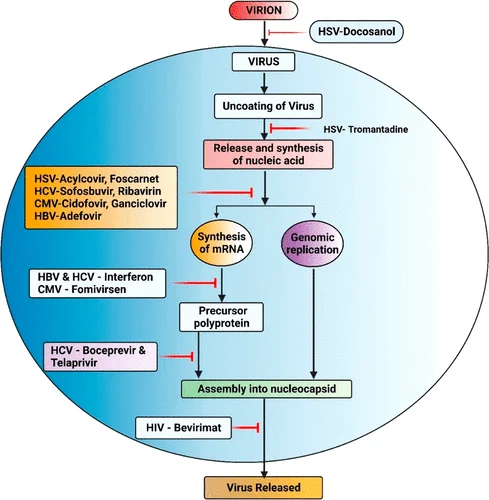
Several findings indicate that Ad12 remained the primary oncogenic driver:
- No tumors developed in animals treated only with MNU.
- Tumors appeared exclusively in virus-inoculated eyes.
- Viral T-antigen was consistently detected within tumor cells.
- Histological features were identical regardless of MNU exposure.
The principal effect of MNU was therefore to accelerate tumor formation by increasing susceptibility of immature retinal precursor cells to viral transformation.
Researchers proposed several possible mechanisms:
- Enhanced vulnerability of fetal neural precursor cells
- Suppression of anti-oncogenic defense pathways
- Increased permissiveness for viral gene expression
- Alteration of cellular differentiation programs
However, the precise interaction between nitrosourea carcinogens and adenoviral oncogenesis remained unresolved.$
Biological Significance of Adenovirus Type 12 T-Antigen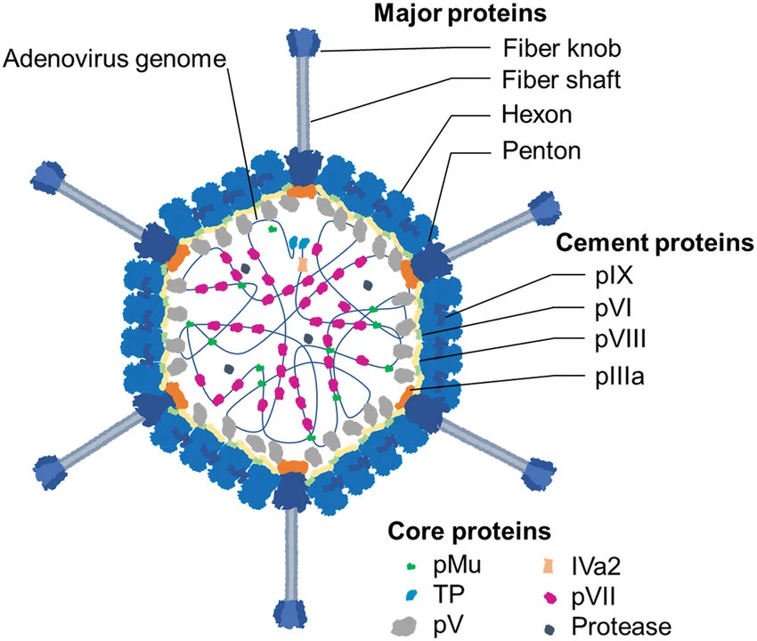
The Ad12 T-antigen represents an early viral protein expressed during infection and transformation. Although its exact biological function remains incompletely understood, it is considered a marker of partial viral genome expression within transformed cells.
The consistent detection of T-antigen in retinal tumor cells strongly supports the conclusion that:
- Viral genes remained active during tumor progression
- The tumors originated directly from adenoviral transformation
- Viral oncogenic pathways contributed to sustained malignant behavior
The intracellular distribution of T-antigen observed in this study closely matched patterns previously described in adenovirus-transformed cells.
Mechanisms of Retinal Tumor Susceptibility
The study also highlighted the exceptional sensitivity of immature retinal precursor cells to oncogenic transformation.
Several factors may explain this vulnerability:
- High proliferative activity of neonatal retinal neuroblasts
- Incomplete cellular differentiation
- Increased permissiveness for viral replication
- Reduced genomic stability during development
The low overall tumor incidence compared with central nervous system tumors likely resulted from:
- Technical limitations of neonatal intraocular injection
- Small vitreous cavity size in newborn rats
- Limited viral inoculum volume
- Smaller populations of susceptible retinal progenitor cells
Nevertheless, the successful induction of retinal tumors represented a major breakthrough in experimental ocular oncology.
Scientific Importance and Future Perspectives
This landmark experimental model provided one of the earliest demonstrations that viral oncogenesis can induce retinal tumors resembling human retinoblastoma.
The findings established several important concepts:
- Immature retinal precursor cells are highly susceptible to oncogenic transformation.
- Viral and chemical carcinogens can cooperate during tumor initiation.
- Adenoviral oncogenesis can target sensory neuroepithelial tissues.
- Experimental retinoblastoma-like tumors can reproduce key features of human disease.
These discoveries contributed significantly to understanding:
- Neuro-oncology
- Viral carcinogenesis
- Retinal developmental biology
- Pediatric ocular tumors
- Mechanisms of neuronal transformation
Today, these early studies remain historically important because they helped pave the way for modern investigations into molecular oncology, viral cancer biology, retinal stem cells, and tumor suppressor pathways involved in human retinoblastoma.